Imaging window nanofabricated by direct laser writing for intravital assessment of biomaterials
Duration 14/01/2018 – 13/07/2021 – Funded by MIUR-FARE2017 – G.A. nr R16ZNN2R9K – Publications are linked at the bottom of this page
VIDEO with project results
Project abstract
New biomaterials must be tested carefully following specific regulations that are based on the histopathological analysis of tissues from experimental animals. Such protocols are ethically undesirable, expensive and subjective. Routine clinical use of bulk biomaterials require the reduction of the costs of these biocompatibility tests, which have become unsustainable for small-medium industries.
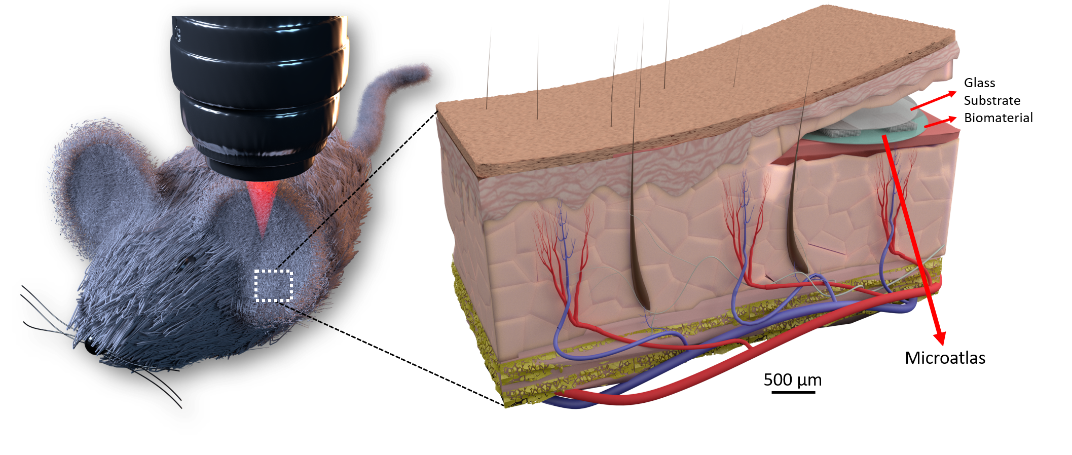
Therefore, I aim at the development of a radically new method and relevant protocols for intravital optical imaging of cells dynamics in response to biomaterial implantation. I will develop a miniaturized chip and I will implant it subcutaneously in transgenic mice expressing fluorescent immune cells. The implanted chip will host a sample of biomaterial and will be colonized in situ by the mouse tissue. The chip, built by two-photon laser polymerization, will contain a micro-scaffold with features of the size in the range 30-50 micrometres and will act as an atlas to reposition the field of view for repeated intravital observations of cells by two-photon fluorescence microscopy.
This technological platform will allow unprecented quantitative and continuous analyses of the host inflammatory response to implants using real time longitudinal studies by intravital imaging of the chips without sacrificing the mice. This project will have a scientific impact on the quantitative analysis of the host reaction to biomaterial implant or, in perspective, to pharmaceutical treatments as well. This will be made possible by the implementation of adaptive optics methods in microscopy, assisted by the use of laser-printed microstructures acting as fluorescent beacons. Its future development will have an ethical impact on reduction of the number of animals sacrificed in biomaterials/drug testing and an economic impact on reduction of the developmental costs of new biomaterials.
Press
Italian Healthcare World, November 10th, 2020
Ansa.it, June 7, 2020
Fanpage.it, June 8, 2020
Giornale di Sicilia, June 7, 2020
Corrierequotidiano.it, June 7, 2020
Aboutpharma.com, June 6, 2020
Insalutenews.it, June 6, 2020
AltoAdige.it, June 6, 2020
Italycom.net, June 6, 2020
Nel Cuore.org, June 6, 2020
Cronachediscienza.it.June 4, 2020
Mynewsdesk.com, June 4, 2020
Eurekaalert.org, June 4, 2020
La Statale News, June 4, 2020
Publications
Conci C, Jacchetti E, Sironi L, Gentili L, Cerullo G, Osellame R, Chirico G, Raimondi MT. A miniaturized imaging window to quantify intravital tissue regeneration within a 3D micro scaffold in longitudinal studies. Adv. Optical Mater. 2022, 2101103. https://doi.org/10.1002/adom.202101103
Musi CA, Colnaghi L, Giani A, Priori EC, Marchini G, Tironi M, Conci C, Cerullo G, Osellame R, Raimondi MT, Remuzzi A, Borsello T. Effect of 3D Synthetic Microscaffold Nichoid on the Morphology of Cultured Hippocampal Neurons and Astrocytes. Cells. 2022 Jun 23;11(13):2008. doi: 10.3390/cells11132008
Raimondi MT, Barzaghini B, Bocconi A, Conci C, Martinelli C, Nardini A, Testa C, Carelli S, Cerullo G, Chirico G, Gottardi R, Osellame R, Remuzzi A, Laganà M, Jacchetti E, Micro structured tools for cell modeling in the fourth dimension, Proc. SPIE 11786, Optical Methods for Inspection, Characterization, and Imaging of Biomaterials V, 117861T (20 June 2021); doi: 10.1117/12.259332
Perottoni S, Neto NGB, Di Nitto C, Dmitriev RI, Raimondi MT, Monaghan MG. Intracellular label-free detection of mesenchymal stem cell metabolism within a perivascular niche-on-a-chip. Lab on a Chip. Feb 2020. doi:10.1039/d0lc01034k.
Parodi V, Jacchetti E, Bresci A, Talone B, Valensise CM, Osellame R, Cerullo G, Polli D and Raimondi MT. Characterization of Mesenchymal Stem Cell Differentiation within Miniaturized 3D Scaffolds through Advanced Microscopy Techniques. Int. J. Mol. Sci. 2020, 21, 8498; doi:10.3390/ijms21228498
Parodi V, Jacchetti E, Osellame R, Cerullo G, Polli D and Raimondi MT. (2020) Nonlinear Optical Microscopy: From Fundamentals to Applications in Live Bioimaging. Front. Bioeng. Biotechnol. 8:585363. doi: 10.3389/fbioe.2020.585363odi
Raimondi MT, Donnaloja F, Barzaghini B, Bocconi A, Conci C, Parodi V, Jacchetti E, Carelli S. Bioengineering tools to speed up the discovery and preclinical testing of vaccines for SARS-CoV-2 and therapeutic agents for COVID-19. Theranostics 2020; 10(16):7034-7052. doi:10.7150/thno.47406. Available from: doi:10.7150/thno.47406
Steimberg, N., Bertero, A., Chiono, V., Dell’Era, P., Di Angelantonio, S., Hartung, T., Perego, S., Raimondi, M.T., Xinaris, C., Caloni, F., De Angelis, I., Alloisio, S. and Baderna, D. (2020) “iPS, organoids and 3D models as advanced tools for in vitro toxicology”, ALTEX – Alternatives to animal experimentation, 37(1), pp. 136-140. DOI https://doi.org/10.14573/altex.1911071
Conci C, Bennati L, Bregoli C, Buccino F, Danielli F, Gallan M, Gjini E, Raimondi MT. Tissue engineering and regenerative medicine strategies for the female breast. J Tissue Eng Regen Med. 2020 Feb;14(2):369-387. doi 10.1002/term.2999
Conci C, Jacchetti E, Zandrini T, Sironi L, Collini M, Chirico G, Cerullo G, Osellame R, Raimondi MT. Quantification of the foreign body reaction by means of a miniaturized imaging window for intravital nonlinear microscopy. Biomed Sci and Eng. 2019; 3:106. doi:10.4081/bse.2019.106

